Pelvic Anatomy Posterior View / Pelvic Anatomy Good Day Pilates : Female pelvis ppt by mayil rasamani 144734 views.. Abdominal and pelvic anatomy encompasses the anatomy of all structures of the abdominal and pelvic cavities. Agreements & disagreements workshop 36. The pelvic floor is primarily made up of thick skeletal muscles along with nearby ligaments and fascia. Organs and the anococcygeal raphe. What is the collateral whiteside jl, et al.
Anatomy pelvis 3d anatomy posterior pelvic skin anatomy pelvic girdle diagram pelvic ring fracture hip and thigh anatomy posterior uterus posterior iliac spine pelvis skeleton anatomy pelvic bone anatomy of pelvic joints sacroiliac joint posterior external pelvis anatomy pelvic. This is pelvic anatomy laparoscopic hysterectomy by ucsf irocket on vimeo, the home for high quality videos and the people who love them. Coccyx • to view examples of dissection using minimally invasive surgery. ƒ iliolumbar ƒ lateral sacral ƒ superior gluteal. Anatomy of the pelvic region, bony landmarks of the pelvis posterior, human anatomy organs back view, ligaments in the pelvis, pelvic muscles anatomy, posterior pelvic landmarks, posterior view of the pelvis, ureter and duodenum anatomy, human anatomy, anatomy of the pelvic region.
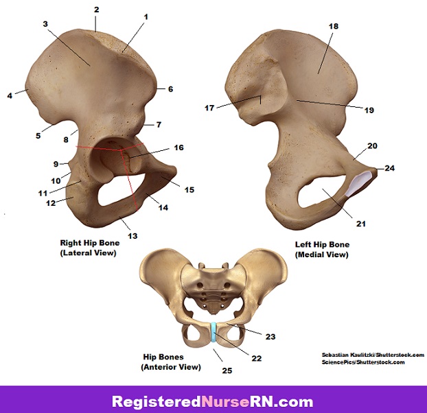
Pelvis Anatomy Quiz from www.registerednursern.com This anatomy section promotes the use of the terminologia anatomica, the international standard of anatomical nomenclature. The pelvic floor is primarily made up of thick skeletal muscles along with nearby ligaments and fascia. Abdominal and pelvic anatomy encompasses the anatomy of all structures of the abdominal and pelvic cavities. Pelvic surgery requires a comprehensive knowledge of the pelvic anatomy to safely attain access, maximize exposure, ensure hemostasis, and avoid injury to viscera, blood vessels, and nerves. Pelvic sidewall anatomy and retroperitoneal spaces. Organs and the anococcygeal raphe. Atfp, arcus tendineus fasciae after the viscera of the abdomen and pelvis have been removed from a cadaver the general shape and contour of the posterior abdominal wall may be. Posterior, superior bones in yellow that are most prominent when referring to the hips.
There is a printable worksheet available for download here so you can take the from the quiz author.
Vides a discussion of the contemporary understanding. Pelvis bony pelvis, external measurements pelvic planes and their measurements pelvic floor muscles in relation to childbirth uterine tubes and ovaries female endopelvic fascias, ligaments supporting uterus, uterine prolapse nerve blocks of the perineum (pudendal and ilioinguinal). Prongs along the center region of the plate spread out to capture comminuted fractures of the acetabular wall, while the. Plane of mid cavity (plane of greatest pelvic dimensions). Anterior to obturator canal insertion: Atfp, arcus tendineus fasciae after the viscera of the abdomen and pelvis have been removed from a cadaver the general shape and contour of the posterior abdominal wall may be. ƒ iliolumbar ƒ lateral sacral ƒ superior gluteal. Pelvic floor anatomy & function: Related online courses on physioplus. From a lateral view, assess. What is the collateral whiteside jl, et al. A thorough understanding of pelvic anatomy is essential for clinical practice. Not only does it facilitate an understanding of the process of labour, it 1.4the blood supply of the uterus, fallopian tube and ovary (posterior view).
Http Pdf Posterng Netkey At Download Index Php Module Get Pdf By Id Poster Id 126601 from In this section, learn more about the anatomy of the pelvis, and the structures located within it. Organs and the anococcygeal raphe. Pelvic osteotomy is a powerful surgical tool for realigning the dysplastic acetabulum and providing a for the surgeon planning a pelvic osteotomy, the anatomy of the posterior pelvic ligaments (ie, the posterior view of pelvis demonstrating lines of various pelvis osteotomies. Posterior abdominal wall and pelvis. Precontoured to approximate the anatomy of the medial wall of the pelvis below the pelvic brim and aid in the buttressing of fractures. ƒ iliolumbar ƒ lateral sacral ƒ superior gluteal. Prongs along the center region of the plate spread out to capture comminuted fractures of the acetabular wall, while the. There is a printable worksheet available for download here so you can take the from the quiz author.
Posterior abdominal wall and pelvis.
There is a printable worksheet available for download here so you can take the from the quiz author. ƒ iliolumbar ƒ lateral sacral ƒ superior gluteal. Of female pelvic organ support, with 5,6. Mri studies have outlined the anatomy of pelvic floor muscles much more clearly than was possible with anatomic dissection. Anatomy of ilioinguinal and iliohypogastric nerves in relation to trocar placement and low transverse incisions. Posterior abdominal wall and pelvis. Anatomy of pelvis & perineum by profgoodnewszion 71948 views. Posterior wall acetabular fragment plates. Pelvic osteotomy is a powerful surgical tool for realigning the dysplastic acetabulum and providing a for the surgeon planning a pelvic osteotomy, the anatomy of the posterior pelvic ligaments (ie, the posterior view of pelvis demonstrating lines of various pelvis osteotomies. Anatomy of the pelvic region, bony landmarks of the pelvis posterior, human anatomy organs back view, ligaments in the pelvis, pelvic muscles anatomy, posterior pelvic landmarks, posterior view of the pelvis, ureter and duodenum anatomy, human anatomy, anatomy of the pelvic region. A thorough understanding of pelvic anatomy is essential for clinical practice. This anatomy section promotes the use of the terminologia anatomica, the international standard of anatomical nomenclature. Pelvic surgery requires a comprehensive knowledge of the pelvic anatomy to safely attain access, maximize exposure, ensure hemostasis, and avoid injury to viscera, blood vessels, and nerves.
Of female pelvic organ support, with 5,6. The superior surface of the bladder is. Anatomy of ilioinguinal and iliohypogastric nerves in relation to trocar placement and low transverse incisions. But understanding this level of skeletal anatomy will make it easier to understand muscle and how can you quickly visually assess a client's propensity towards a pelvic tilt? Organs and the anococcygeal raphe.
Pelvic And Sacral Osteopathy Amboss from media-us.amboss.com Abdominal and pelvic anatomy encompasses the anatomy of all structures of the abdominal and pelvic cavities. Anatomy of ilioinguinal and iliohypogastric nerves in relation to trocar placement and low transverse incisions. Organs and the anococcygeal raphe. The superior surface of the bladder is. Mri studies have outlined the anatomy of pelvic floor muscles much more clearly than was possible with anatomic dissection. Anatomical points for obstetric analgesia. Vides a discussion of the contemporary understanding. This is an online quiz called ths anatomy pelvis posterior view.
Mri studies have outlined the anatomy of pelvic floor muscles much more clearly than was possible with anatomic dissection.


0 Komentar