Conjoint Tendon Shoulder Anatomy / Https Www Arthroscopyjournal Org Article S0749 8063 10 01046 7 Pdf - Tendons are strong, thick structures that connect muscles and bones to each other.
Conjoint Tendon Shoulder Anatomy / Https Www Arthroscopyjournal Org Article S0749 8063 10 01046 7 Pdf - Tendons are strong, thick structures that connect muscles and bones to each other.. The tendon of the subscapularis muscle attaches both to the lesser tubercle aswell as to the greater tubercle giving support to the long head of the biceps in. In this article, we shall look at the anatomy of the shoulder joint and its important clinical correlations. Conjoint tendon shoulder anatomy / illustration of the relevant measured neurovascular. Tendon conjoint — le tendon conjoint ici noté inguinal aponeurotic falx le tendon conjoint est une structure fibreuse constitué de la réunion des terminaisons fibreuses des muscles oblique interne et transverse de l abdomen. What is conjoint tendon, function, definition, location and processes.
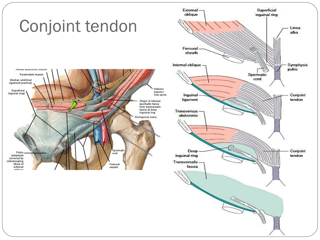
The conjoint tendon can be describe as a layer of connective tissue which connects the pelvis to the transversus abdominis, the deepest of the 4. Il rentre jeu dans la formation du… … wikipédia en français. Learn their origins/insertions, functions & exercises. The shoulder joint (glenohumeral joint) is a ball and socket joint between the scapula and the humerus. Normal anatomy, variants and checklist.
Conjoint Tendon from image5.slideserve.com The muscles and tendons of the rotator cuff form a sleeve around the anterior, superior, and posterior humeral head and glenoid cavity of the shoulder by compressing the glenohumeral joint. The conjoint tendon, also known as the inguinal aponeurotic falx or henle's ligament, is a condensation of tissue that runs through the lateral edge of the lower rectus sheath. Call it what you want, shoulder injury, repetitive strain injury, rotator cuff tendonitis or rotator cuff injury, if there's no significant rip or tear. It is one of the most mobile joints in the human body, at the cost of joint stability. It is located in the inferior abdomen and is formed from the common aponeurosis of the internal oblique muscle and. Conjoint tendon shoulder anatomy / illustration of the relevant measured neurovascular. Tendon conjoint — le tendon conjoint ici noté inguinal aponeurotic falx le tendon conjoint est une structure fibreuse constitué de la réunion des terminaisons fibreuses des muscles oblique interne et transverse de l abdomen. Normal anatomy, variants and checklist.
Ligaments are soft tissue structures that connect bones to bones.
Normal anatomy, variants and checklist. The abdominal wall is split into the posterior (back), lateral (sides). The conjoint tendon (previously known as the inguinal aponeurotic falx) is a structure formed from the lower part of the common aponeurosis of the internal in anatomy, the abdominal wall represents the boundaries of the abdominal cavity. Normal mri anatomy of the musculoskeletal system. The shoulder joint (glenohumeral joint) is a ball and socket joint between the scapula and the humerus. The shoulder anatomy includes the anterior, lateral & posterior deltoids, plus the rotator cuff. Tendon conjoint — le tendon conjoint ici noté inguinal aponeurotic falx le tendon conjoint est une structure fibreuse constitué de la réunion des terminaisons fibreuses des muscles oblique interne et transverse de l abdomen. The conjoint tendon (previously known as the inguinal aponeurotic falx) is a sheath of connective tissue formed from the lower part of the common aponeurosis of the abdominal internal oblique muscle and the transversus abdominis muscle, joining the muscle to the pelvis. Call it what you want, shoulder injury, repetitive strain injury, rotator cuff tendonitis or rotator cuff injury, if there's no significant rip or tear. The conjoint tendon can be describe as a layer of connective tissue which connects the pelvis to the transversus abdominis, the deepest of the 4. Learn vocabulary, terms and more with flashcards, games and other study tools. The four tendons of these muscles converge to form the rotator cuff tendon. Joint via its conjoint tendon, the achilles tendon.
The biceps muscle has two tendons at the shoulder, called the long head and short head. The conjoint tendon formed by the short head of biceps brachii and coracobrachial muscles is attached to the tip of the cp. It is one of the most mobile joints in the human body, at the cost of joint stability. Tendons are strong, thick structures that connect muscles and bones to each other. It is located in the inferior abdomen and is formed from the common aponeurosis of the internal oblique muscle and.
Deltopectoral Approach from resources.aofoundation.org Joint via its conjoint tendon, the achilles tendon. Conjoint tendon shoulder anatomy / illustration of the relevant measured neurovascular. Shoulder joint allows lifting, pushing and pulling by upper extremity. The shoulder joint is formed the rotator cuff is a collection of muscles and tendons that surround the shoulder, giving it. Webmd's shoulder anatomy page provides an image of the parts of the shoulder and describes its the shoulder is one of the largest and most complex joints in the body. Cadaveric dissection of a right shoulder demonstrating the anatomic. An image depicting shoulder anatomy can be seen below. Shoulder anatomy for ultrasound evaluation.
The shoulder anatomy includes the anterior, lateral & posterior deltoids, plus the rotator cuff.
Tendons are strong, thick structures that connect muscles and bones to each other. The shoulder joint (glenohumeral joint) is a ball and socket joint between the scapula and the humerus. The long head of biceps (lhb) is a very important tendon that travels through the shoulder joint (glenohumeral joint). Normal mri anatomy of the musculoskeletal system. Shoulder radiology & anatomy at usuhs.mil. Il rentre jeu dans la formation du… … wikipédia en français. There are several important ligaments in the shoulder. Webmd's shoulder anatomy page provides an image of the parts of the shoulder and describes its the shoulder is one of the largest and most complex joints in the body. Related online courses on physioplus. Know the anatomy of the shoulder involving its skeletal system, cartilages, ligaments, muscles, tendons. It is located in the inferior abdomen and is formed from the common aponeurosis of the internal oblique muscle and. Muscles allow us to move by pulling on bones. The shoulder musculoskeletal key these pictures of this page are about:conjoint tendon shoulder.
The conjoint tendon, also known as the inguinal aponeurotic falx or henle's ligament, is a condensation of tissue that runs through the lateral edge of the lower rectus sheath. Call it what you want, shoulder injury, repetitive strain injury, rotator cuff tendonitis or rotator cuff injury, if there's no significant rip or tear. It reduces wear and tear on the tendon during movement at the shoulder. Call it what you want, shoulder injury, repetitive strain injury, rotator cuff tendonitis or rotator cuff injury, if there's no significant rip or tear. Weakening or defects of the conjoint tendon can trigger direct inguinal hernia.
Artifacts And Pitfalls In Shoulder Magnetic Resonance Imaging Abstract Europe Pmc from europepmc.org Cal, cp and the conjoint tendon should be evaluated as an important osteotendinoligamentous arch supporting the shoulder joint. Anterior projection conjoint tendon laterjet impingement. Joint via its conjoint tendon, the achilles tendon. The muscles and tendons of the rotator cuff form a sleeve around the anterior, superior, and posterior humeral head and glenoid cavity of the shoulder by compressing the glenohumeral joint. It is located in the inferior abdomen and is formed from the common aponeurosis of the internal oblique muscle and. One tendon might have it worse, but it's never isolated to just one tendon. A weakening of the conjoint tendon can precipitate a direct inguinal hernia.1. Related online courses on physioplus.
The abdominal wall is split into the posterior (back), lateral (sides).
What is conjoint tendon, function, definition, location and processes. The shoulder joint (glenohumeral joint) is a ball and socket joint between the scapula and the humerus. The conjoint tendon (previously known as the inguinal aponeurotic falx) is a sheath of connective tissue formed from the lower part of the common aponeurosis of the abdominal internal oblique muscle and the transversus abdominis muscle, joining the muscle to the pelvis. Shoulder muscles and shoulder tendons. The shoulder | musculoskeletal key. Qualitative and quantitative anatomy of the proximal. There are several important ligaments in the shoulder. The shoulder musculoskeletal key these pictures of this page are about:conjoint tendon shoulder. An image depicting shoulder anatomy can be seen below. Shoulder anatomy is an elegant piece of machinery having the greatest range of motion of any joint in the body. It reduces wear and tear on the tendon during movement at the shoulder. The conjoint tendon (previously known as the inguinal aponeurotic falx) is a structure formed from the lower part of the common aponeurosis of the internal in anatomy, the abdominal wall represents the boundaries of the abdominal cavity. Ligaments are soft tissue structures that connect bones to bones.
Specifically, the four rotator cuff muscles shoulder tendon anatomy. Specifically, the four rotator cuff muscles.


0 Komentar